CIVA-RX – Rayo X
Hacedor:CEA
Radiography testing simulation allows to easily do feasibility studies on radiographic images obtained for a given object, under various angles, and by using various radiation sources. The software is easy to use and allows rapid definition of all relevant parameters, such as part definition and positioning, source and detector selection and positioning, insertion of various kinds of flaws, and calculation options.
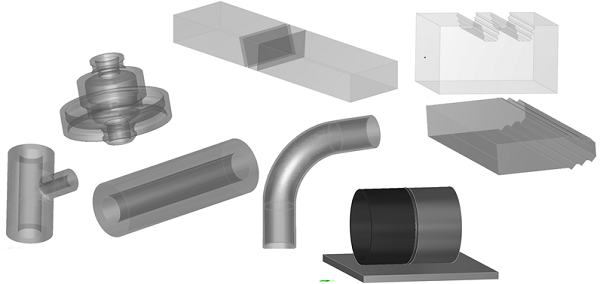
Specimen definition
- Geometries: Canonical (flat, cylindrical, conical), pre-set components (nozzle, butt or tee weld, turbine blade, elbow), import of 2D and 3D CAD files
- Materials: Materials library with over 110 elements and alloys available. User defined alloys can be added. Homogeneous and heterogeneous (more than one material) components can be defined
Radiography sources and detectors
CIVA is prepared for simulating all practically relevant radiographic sources and detectors:
- X-ray sources: The source’s spectrum and intensity can by defined, by either using a predefined spectrum, loading an experimental spectrum, or using a spectrum calculator based on physical parameters of the source.
- Gamma-ray sources: The radioisotope and its activity need to be defined. Classical sources are pre-defined (Co 60, Ir 192, Se 75), other sources can be added.
- High energy sources: Library of high energy spectra available (linear accelerator and betatrons)
- Detector types: Silver films, image plates of high sensitivity or resolution, digital detectors (flat panels), CCD scintillators
- Detector parameters: Shape (flat or curved), filters present or not, noise and granularity
Radiography inspection simulation
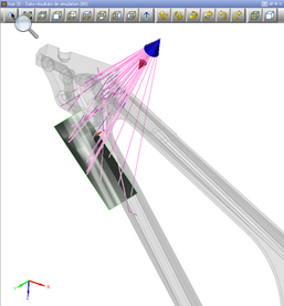
CIVA simulates the interaction of the radiographic beam with the sample, and the subsequent detection of the resulting radiographic field with the detector.
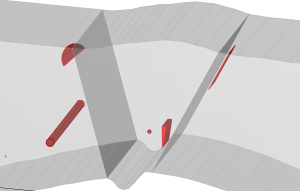
 Defect types: Canonical (planar, spherical, ellipsoidal, trapezoidal), or import of 3D CAD geometries
- Flaw materials: Can be selected from the same list as the sample material
- Defect number: Single defect, multiple defects
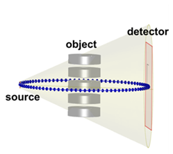
- Acquisition modes: 2D radiography: Use of analytical Beer-Lambert and Monte-Carlo simulation. 3D CT-scanning: Circular or helical scanning available.